
File:Carbon nanotubes penetrating lung cell.tif

此TIF文件的JPG预览的大小:800 × 554像素。 其他分辨率:320 × 222像素 | 640 × 443像素 | 1,024 × 709像素 | 1,280 × 886像素。
原始文件 (1,280 × 886像素,文件大小:1.1 MB,MIME类型:image/tiff)
摘要
| 描述 |
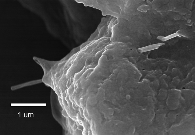
English: Figure 4: Field emission scanning electron microscope (FESEM) image of multiwalled carbon nanotube (MWCNT) penetration of alveolar epithelial cells. As shown in this micrograph, MWCNT were rapidly incorporated into the alveolar epithelium. Micrograph shows two MWCNT passing through an alveolar epithelial cell 1 day after aspiration (80 ug dose). The two fibers are running together on the right side of the cell and appear to separate within the cell as they pass out on the left side of the cell. Another MWCNT penetrates the epithelium in the upper right of micrograph. |
| 日期 | |
| 来源 | Distribution and persistence of pleural penetrations by multi-walled carbon nanotubes |
| 作者 | Robert R. Mercer, Ann F. Hubbs, James F. Scabilloni, Liying Wang, Lori A. Battelli, Diane Schwegler-Berry, Vincent Castranova and Dale W. Porter / NIOSH |
许可协议
|
|
文件历史
点击某个日期/时间查看对应时刻的文件。
| 日期/时间 | 缩略图 | 大小 | 用户 | 备注 | |
|---|---|---|---|---|---|
| 当前 | 2016年7月15日 (五) 20:18 |  | 1,280 × 886(1.1 MB) | Antony-22 | User created page with UploadWizard |
文件用途
以下页面使用本文件:
全域文件用途
以下其他wiki使用此文件:
- en.wikipedia.org上的用途
- Carbon nanotube
- Nanotoxicology
- Toxicology of carbon nanomaterials
- Health and safety hazards of nanomaterials
- Template:Did you know nominations/Health and safety hazards of nanomaterials
- User:Antony-22/Gallery/Others
- Wikipedia:Meetup/Cincinnati/NIOSH 2017
- Wikipedia:Meetup/Columbus/Emerging technologies Edit-a-thon
- User:John P. Sadowski (NIOSH)/National Nanotechnology Day 2019
- Brain health and pollution
- Wikipedia:Meetup/Cincinnati/Emerging Technologies Edit-a-thon 2022
- fa.wikipedia.org上的用途
- hu.wikipedia.org上的用途
- pt.wikipedia.org上的用途
- sl.wikipedia.org上的用途